Published Alex Hodgson on February 18, 2019
Part 3.5 – Snapshot of a Flow Cell Reaction
All you I Spy aficionados may have noticed something peculiar in one of the CDO-PTAD spectra in Part 3 namely 1,3-cyclohexadiene-PTAD. Its spectrum has several spectral “fingers” that the other spectra do not. Why is that? Let’s put on our organic chemistry caps and dig out those notes on reaction mechanisms. (DISCLAIMER: We cannot be held responsible for any traumatic flashbacks to your college days. We all had that one scarring class.)
Doing a quick perusal through the VUV spectra library, we find the “fingers” of our adduct spectrum line up perfectly with the “fingers” of carbon monoxide’s spectrum (Figure 1). This means that unless somehow the intact 1,3-cyclohexadiene-PTAD adduct happens to have this exact same feature, we’re seeing a “coelution” of the adduct (in some form) with carbon monoxide.
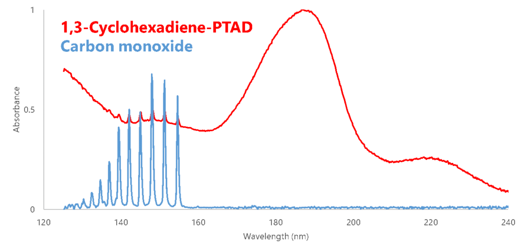
Figure 1. Overlaying the VUV spectra for 1,3-cyclohexadiene-PTAD and carbon monoxide, it looks to be a perfect match for the spectral “fingers” for each compound.
So where is that CO coming from? If we look at the structure of the CDO-PTAD adduct in Figure 2, we can envision a reaction mechanism by which we kick out a CO molecule; what the resulting structure would be is difficult to tell. Running the same sample on a GC-MS, the mass spectrum we get for 1,3-cyclohexadiene-PTAD shows molecular ion at 255 Da and an intense fragment ion at 227 that represents the loss of CO (Figure 3).
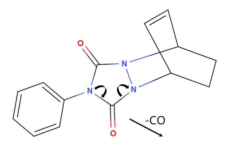
Figure 2. A potential mechanism for a flow cell/ion source reaction that would explain the loss of CO in both the VUV and mass spectra.
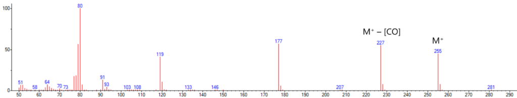
Figure 3. The mass spectrum of the same 1,3-cyclohexadiene-PTAD peak confirms a loss of CO in the detector itself, as we see response for both M+ and M+ – [CO] ions.
Stumbled across any interesting mystery features in your VUV spectra? Let us know in the comments!
Author

Alex Hodgson
Alex Hodgson is an Applications Chemist at VUV Analytics, Inc. His current research focuses on vacuum ultraviolet applications for the flavor and fragrance industries, among a variety of other VUV-related endeavors. Prior to coming to VUV Analytics, Alex was a member of the Center for Disease Control’s team in Atlanta, where his research was on tobacco exposure biomarkers. He earned a B.S. in biochemistry from the University of Texas at Austin and an M.S. in biochemistry at the Georgia Institute of Technology.
Related Reading
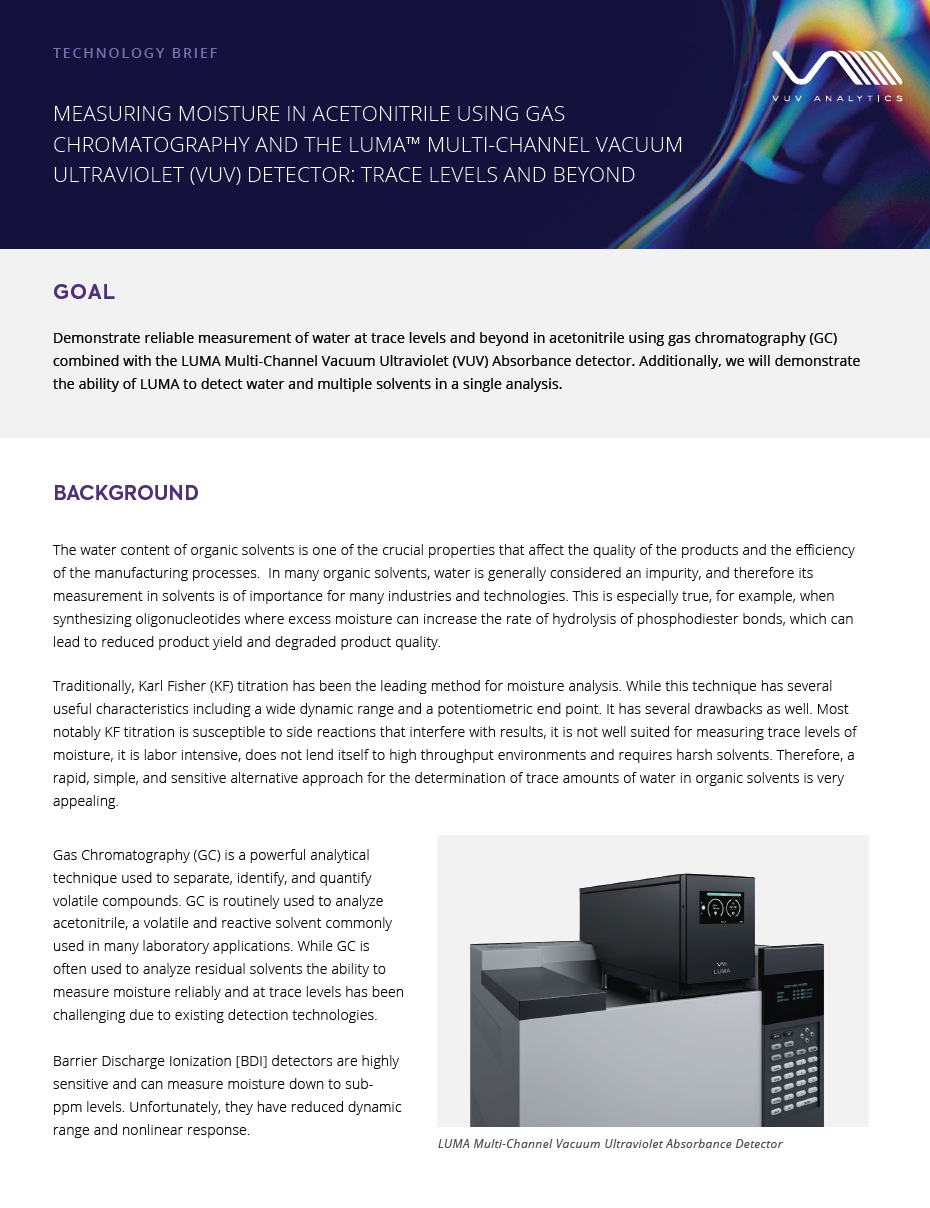
Measuring Moisture in Acetonitrile Using Gas Chromatography and the LUMA Vacuum Ultraviolet (VUV) Detector: Trace Levels and Beyond
The water content of organic solvents is one of the crucial properties that affect the…
Read More >
Standardizing Your Analysis: ASTM D8267 Standards
by Chris Cook
In this blog, we discuss the importance of all standards used in our ASTM D8267…
Read More >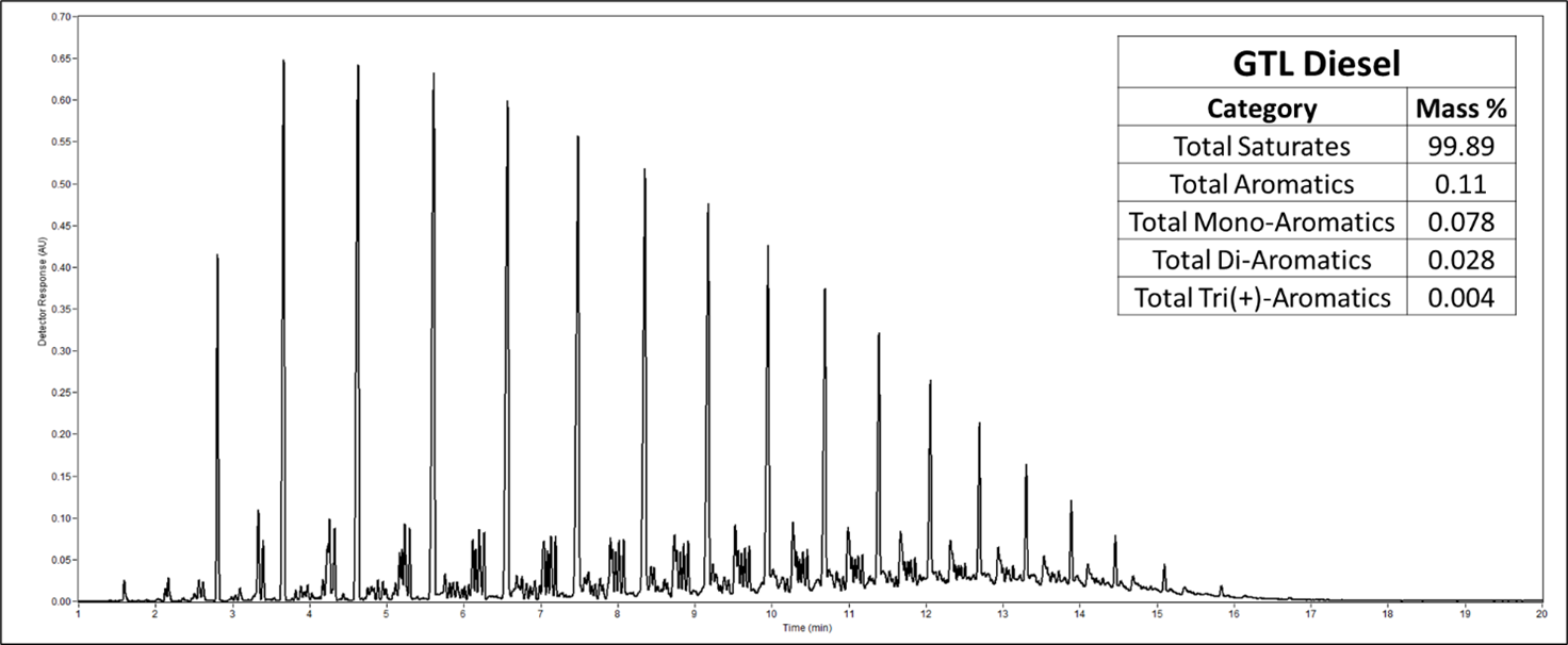
An Update on Our Diesel Method
Alex Hodgson
This blog provides an update to our upcoming diesel analysis method.
Read More >


Leave a Reply